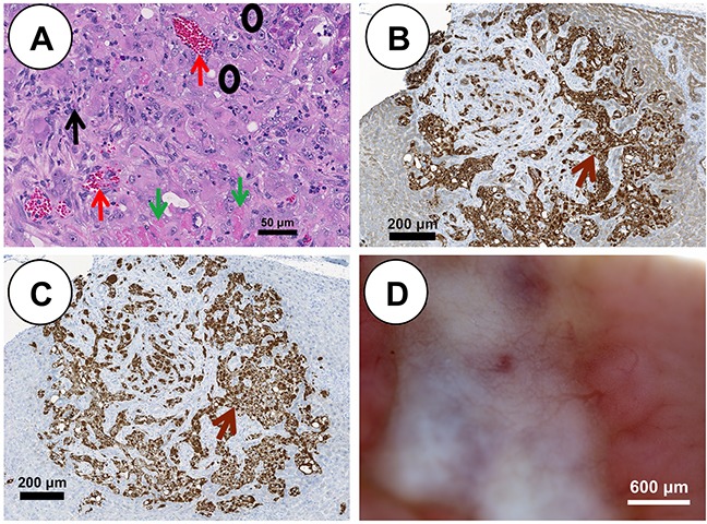
Figure 3. pHCC intrahepatic xenografted tumors recapitulate cytologic and histologic features of human HCC.

(A) H&E stained pHCC intrahepatic xenografted tumor reveals human HCC characteristics including blood vessels (red arrows), stroma (black arrow), neoplastic cells (black circles) and necrotic cells (green arrows) at 21 days post injection (scale bar 50 μm). (B) Positive cytokeratin immunostaining (brown arrow) of pHCC intrahepatic xenografted tumor (scale bar 200 μm). (C) Positive vimentin immunostaining (brown arrow) of pHCC intrahepatic xenografted tumor (scale bar 200 μm). (D) Blood vessel development in pHCC intrahepatic xenografted tumor (scale bar 600 μm).
