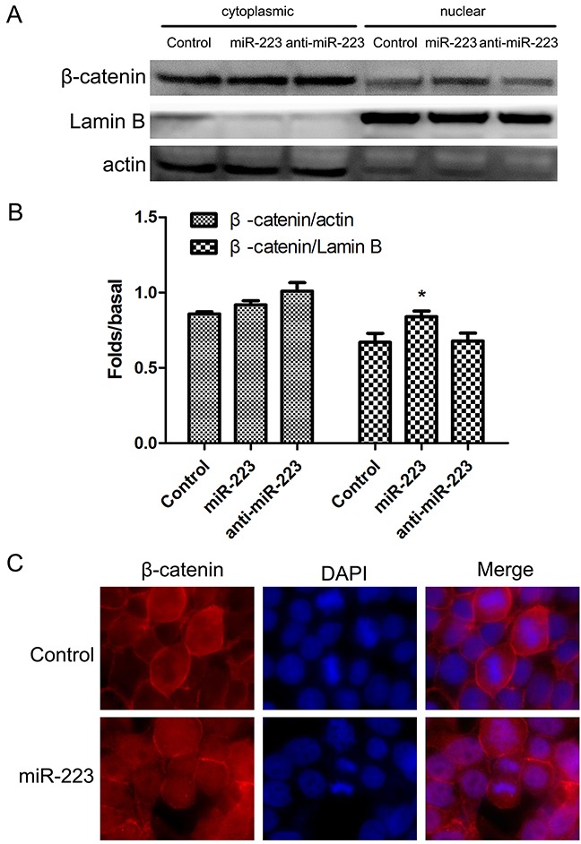
Figure 7. miR-223 overexpression increases nuclear β-catenin in LoVo cells.

LoVo cells were transfected with miR-223 mimics, miR-223 inhibitor, or negative control for 48 h, and cytoplasmic and nuclear β-catenin were detected by western blotting (A). Actin and lamin B were used as cytoplasmic and nuclear protein loading controls, respectively. Representative images shown are from independent experiments repeated three times. Quantified western blotting results (B). n=3; *P<0.05 vs. the control group. LoVo cells were transfected with miR-223 or negative control for 24 h, fixed with 4% paraformaldehyde, and fluorescently stained for β-catenin. DAPI was used to stain nuclei (C). At least five fields of view were imaged using a laser confocal microscope under each condition.
