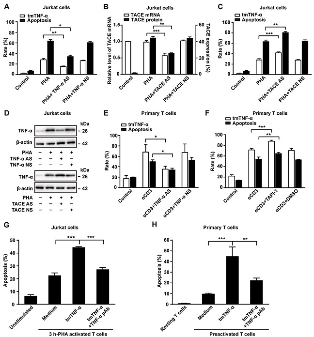
Figure 2. tmTNF-α is involved in AICD.

(A, B, C, D) Jurkat cells transfected with 10 μM of TNF-α AS, TACE AS or NS for 48 h were treated with PHA-P (5 μg/ml) for 24 h. Apoptosis rate (A,C) was evaluated by Annexin V/PI and expression of tmTNF-α (A,C) and TACE (B) on the cell surface was analyzed by flow cytometry. Relative levels of TACE mRNA was detected by Real time qPCR (B). Western blot analysis of tmTNF-α expression (D). (E, F) PHA-preactivated primary T cells were transfected with 10 μM of TNF-α AS or NS for 48 h, and then treated with αCD3 (10 μg/ml) for 24 h. A TACE inhibitor TAPI-1 (10 μM) was added simultaneously with αCD3 treatment, and vehicle DMSO served as a control. Apoptosis was evaluated by Annexin V/PI and tmTNF-α expression was detected by flow cytometry. (G, H) tmTNF-α overexpressing 24 h-PHA activated and fixed Jurkat cells were cocultured with 3 h-PHA activated Jurkat cells at an effector/target ratio of 10:1 for 48 h (G). For primary T cells, tmTNF-α overexpressing, αCD3-restimulated and fixed T cells were co-cultured with PHA-preactivated T cells (preactivated T) at an effector/target ratio of 10:1 for 48h (H). For neutralization of tmTNF-α, effector cells were treated with TNF-α pAb for 30 min and then washed prior to the addition to the target cells. Apoptosis was determined by the Annexin V/PI. All the quantitative data represent means ± S.D. of at least three independent experiments. *p<0.05, **p<0.01, ***p<0.001.
