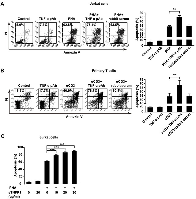
Figure 4. The reverse signaling of tmTNF-α enhances AICD.

(A, C) Jurkat cells were incubated with PHA-P (5 μg/ml), TNF-α pAb (1:100), sTNFR1 or with both PHA-P and TNF-α pAb, or PHA-P and sTNFR1 in indicated concentrations for 24 h. Apoptosis was detected by Annexin V/PI. (B) PHA preactivated primary T cells were stimulated with αCD3 (10 μg/ml), TNF-α pAb, or both for 24 h. Apoptosis was detected by Annexin V/PI. Rabbit serum served as a control. All the quantitative data represent means ± S.D. of at least three independent experiments. **p<0.01, ***p<0.001.
