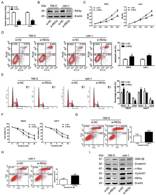
Figure 6. Knockdown of REGγ inhibited proliferation and increased chemosensitivity to sorafenib by suppressing the wnt/β-catenin pathway in RCC cells.

(A and B) 786-O and caki-1 cells were transfected with si-REGγ or si-NC. The relative mRNA levels (A) and the protein expression (B) of REGγ were determined by qRT-PCR and Western blot analysis, respectively. (C) The proliferation of 786-O and caki-1 cells was analyzed by the MTT assay following REGγ knockdown. (D) The apoptosis rate of 786-O and caki-1 cells was determined by flow cytometry following REGγ knockdown. (E) The cell cycle distribution of 786-O and caki-1 cells was measured by flow cytometry following REGγ knockdown. (F) 786-O and caki-1 cells were transfected with si- REGγ or si-NC, and further cultured with sorafenib at various concentrations of 0, 2.5, 5, 10, 20 μM. Following 24 h of sorafenib treatment, cell viability was determined by the MTT assay. (G and H) 786-O and caki-1 cells were transfected with si-REGγ or si-NC, further treated with sorafenib at the concentration of 10 μM for 24 h, and apoptosis was measured by flow cytometry. (I) Western blot measurement of GSK-3β, β-catenin, c-Myc and Cyclin D1 protein expression levels following knockdown of REGγ in 2 RCC cells. β-actin was used as an internal control. The data are presented as mean ± SD of 3 independent experiments. *P < 0.05
