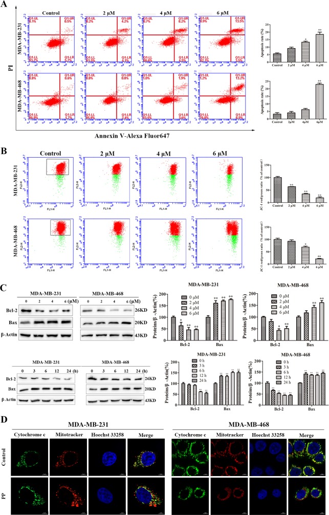
Figure 3. PP induced mitochondrial dysfunction in triple-negative breast cancer cells.
(A) The rates of apoptotic MDA-MB-231 and MDA-MB-468 cells after treatment with PP for 24 h, as determined by Annexin V-Alexa Fluor 647 and PI staining. (B) The mitochondrial membrane potential of MDA-MB-231 and MDA-MB-468 cells treated with PP for 24 h, as measured by flow cytometry with JC-1 staining. (C) The expressions of Bax and Bcl-2 in MDA-MB-231 and MDA-MB-468 cells after treatment with various concentrations of PP for 24 h and 6 μM PP for different periods. (D) MDA-MB-231 and MDA-MB-468 cells were treated with 6 μM PP for 24 h, and their immunofluorescence was assessed. Green: FITC-labeled Cytochrome c; Red: Mito-Tracker-labeled mitochondria; Blue: Hoechst 33258-labeled nuclei. Scale bars = 5 μm. The results were similar in at least three independent experiments. *p < 0.05, **p < 0.01, vs. control group.

