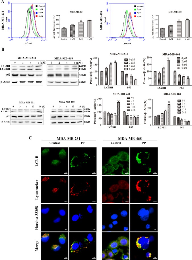
Figure 5. PP promoted the formation of autophagosome in triple-negative breast cancer cells.
(A) MDA-MB-231 and MDA-MB-468 cells were treated with different concentrations of PP for 24 h, stained with AO and analyzed by flow cytometry. (B) Expression levels of LC3BII and p62 by Western blotting in MDA-MB-231 and MDA-MB-468 cells treated with different concentrations of PP and 6 μM PP for different periods. (C) MDA-MB-231 and MDA-MB-468 cells were treated with 6 μM PP for 18 h ad analyzed by immunofluorescence. Green: FITC-labeled LC3B; Red: Lyso-Tracker-labeled lysosomes; Blue: Hoechst 33258-labeled nuclei. Scale bars = 5 μm. The results were similar in at least three independent experiments. *p < 0.05, **p < 0.01, vs. control group.

