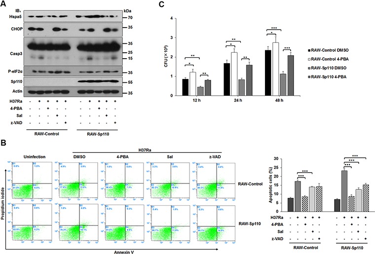
Figure 4. Effect of ER stress response on Sp110-mediated macrophage resistance to Mtb.

(A) Expression of ER stress and apoptotic markers of RAW-Control and RAW-Sp110 cells in response to Mtb infection and chemical inhibitors. RAW-Control and RAW-Sp110 cells were pretreated with 4-PBA, Sal, or z-VAD-FMK for 1 h, and then infected with H37Ra (MOI 5:1) for 24 h. Protein levels of CHOP, Hspa5, Casp9, Casp3, P-eIF2α, and Sp110 were examined by immunoblotting. (B) RAW-Control and RAW-Sp110 cells were pretreated with 4-PBA, Sal, or z-VAD-FMK for 1 h, and then infected with H37Ra (MOI 5:1) for 24 h. Cell apoptosis was quantified by Annexin-V staining followed by flow cytometric analysis. (C) Effect of Sp110-mediated ER stress response on intracellular survival of Mtb. RAW-Control and RAW-Sp110 cells were pretreated with 4-PBA for 1 h and then infected with H37Ra (MOI 5:1) for 4 h. 4-PBA remained for the rest of the infection. The cells were collected at 12 h, 24 h and 48 h post infection with Mtb, and bacteria number was determined by CFU counting. Data are present as means ±SD of three independent experiments,* p< 0.05, **p< 0.01, and *** p< 0.001.
