
Figure 4.
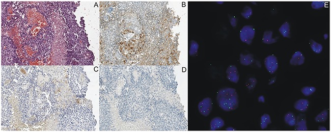
Representative IHC staining of ALK expression using D5F3 antibody and PD-L1 expression using 22C3 and SP142 antibodies on tumor cells in the same NSCLC patients with ALK gene rearrangement detected in FISH (E) and in RT-PCR method. (A) Routine histopathological staining with hematoxylin and eosine. (B) Percentage of tumor cells with abnormal ALK protein expression was 80%. (C and D) Lack of PD-L1 expression in IHC staining using 22C3 and SP142 antibodies.