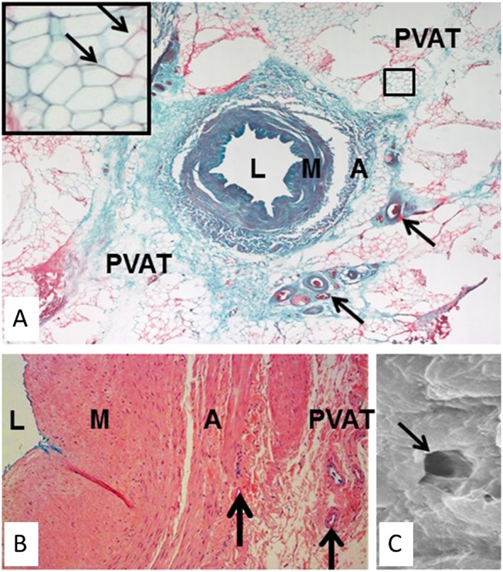
Figure 3.

Inside‐out/outside‐in connections in human no‐touch saphenous vein (SV). (A) Transverse section of a no‐touch SV. Arrows indicate vasa vasorum. Inset, higher magnification of area in (B) showing adipocytes with embedded capillaries (arrows). (B) Part of a transverse section of no‐touch SV perfused with Indian ink (blue stain). There is staining of the lumen and vasa vasorum within the adventitia and PVAT. (C) Scanning electron micrograph of the endothelial surface of the lumen of a no‐touch SV showing the termination of a vasa vasorum (arrow). L, lumen; M, media; A, adventitia. Stained section in (B) provided by Dr Craig Daly (www.cardiovascular.org). Indian ink perfused vein in (C) is from Dr Mats Dreifaldt (unpublished). Scanning electron micrograph in (C) is from Vasilakis et al., 2004.
