
Figure 3.
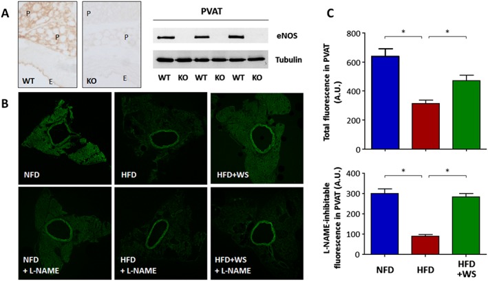
WS® 1442 improves PVAT NO production in HFD mice. (A) eNOS immunohistochemistry staining and western blot analyses were performed using PVAT‐containing aorta samples from C57BL/6J wild‐type mice (WT) or global eNOS knockout mice (KO). E and P indicate endothelium and PVAT respectively. (B) Male C57BL/6J mice were put on a NFD or HFD for 22 weeks starting at the age of 8 weeks. A subgroup of HFD animals were treated with WS® 1442, p.o., during the last 4 weeks of HFD feeding. NO production in PVAT was determined by DAF‐2 DA staining in the absence or presence of the NOS inhibitor L‐NAME. The confocal images shown are representative for five independent experiments with similar results. (C) Shows the quantification fluorescence intensity in PVAT. *P < 0.05, n = 5.