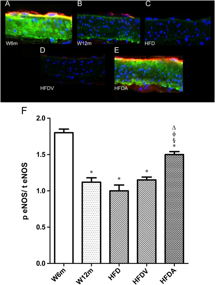
Figure 4.

Effects of HFD and adiponectin treatment on mesenteric eNOS levels in W12m rats, compared with W6m rats. Representative mesenteric sections demonstrating decreased p‐eNOS staining in W12m, HFD and HFDV arteries (B–D). Panel presents mesenteric arteries from W6m (A), W12m (B), HFD (C), HFDV (D) and HFDA (E) groups of rats. Panel (F) contains quantification of the red (p eNOS) to green fluorescence (total eNOS;t eNOS) ratio in the different groups of arteries. Data are mean ± SE (n = 11 animals per group). *P < 0.05, significantly different from W6m group; § P < 0.05, significantly different from W12m group; φ P < 0.05, significantly different from HFD group; Δ P < 0.05, significantly different from HFDV group.
