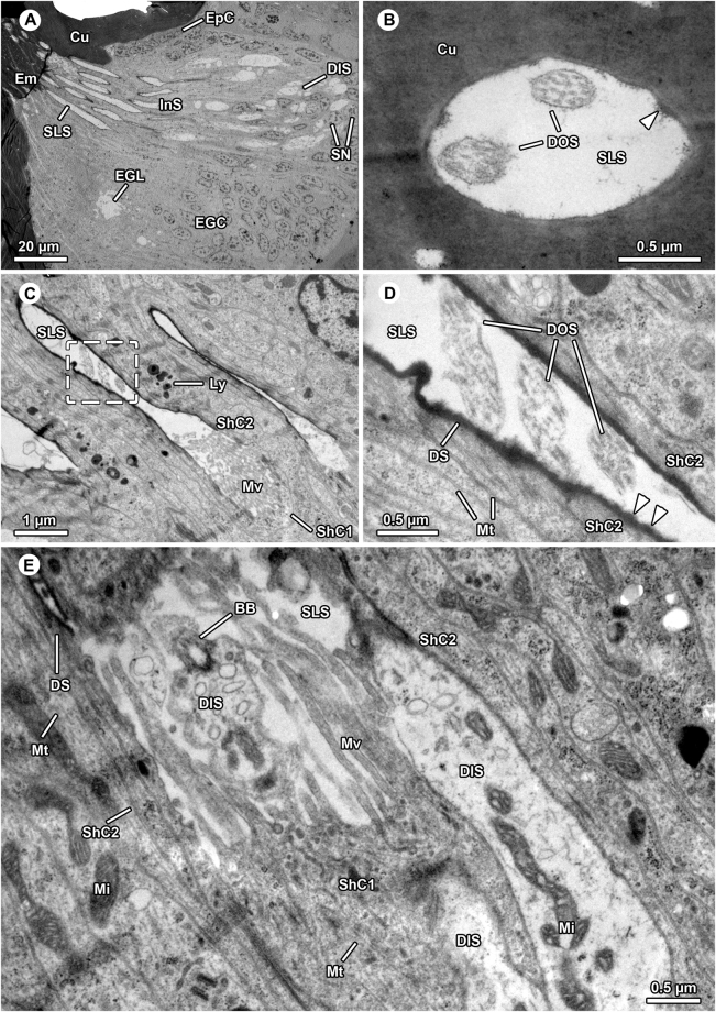
Figure 3.
Ultrastructure of aggregated internalized sensilla in the bulbus of Philodromus cespitum. (A) Overview of distal sensilla components comprising dendrites and sensillum lymph spaces, note that part of embolus gland tissue surrounds the sensilla. (B) Most distal, endocuticular part of sensillum penetrating embolus cuticle, electron-lucent sensillum lymph space bears two dendritic outer segments. The aggregation of fibrillous material are potential remains of the dendritic sheath (arrowhead). (C–D). In the proximal region the dendritic outer segments project through sensillum lymph space which is lined by highly electron-dense dendritic sheaths (C,D) close-up of sector boxed in (C), sensillum lymph space contains three outer dendritic segments in oblique view. Arrowheads point at knob-like protrusions of the dendritic sheath. (E). Transition zone between outer and inner dendritic segments (basal body region), one basal body is visible. The three dendritic inner segments are encompassed by innermost (type-1) sheath cell projecting elongated microvilli into sensillum lymph space. BB – basal body, Cu – cuticle, DIS – dendritic inner segment, DOS – dendritic outer segment, DS – dendritic sheath, Em – embolus, EGC – embolus gland cells, EGL – embolus gland lumen EpC – epidermal cells, InS – internalized sensilla, Ly – lysozomes, Mi – mitochondrium, Mt – microtubuli, Mv – microvilli, SLS – sensillum lymph space, ShC – sheath cell, SN – sheath cell nuclei.

