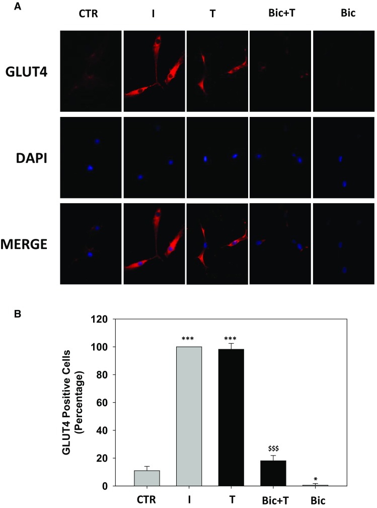
Fig. 4.

Effect of bicalutamide pre-treatment on testosterone-induced GLUT4 translocation in Hfsmc. Immunofluorescence analysis (a) revealed no signal for GLUT4 membrane expression in control (ctr) untreated Hfsmc; positive staining for GLUT4 was observed after 30-min incubation with I (100 nM) or T (100 nM) (upper panels). 1-h pre-treatment with Bic 100 nM completely counteracted T-induced GLUT4 translocation. Cells were incubated with antibody probes specific for GLUT4, followed by incubations with fluorescent secondary antibody. Middle panels represent DAPI blue staining of nuclei; lower panels depict GLUT4/DAPI staining merge. Pictures are representative. Results are derived from four separate experiments, using distinct cell preparations. Percentage of GLUT4 positive cells (*P < 0.05, ***P < 0.001 vs. ctr and $$$ P < 0.001 vs. T) are represented in b. Data are expressed as mean ± SE
