Figure 3.
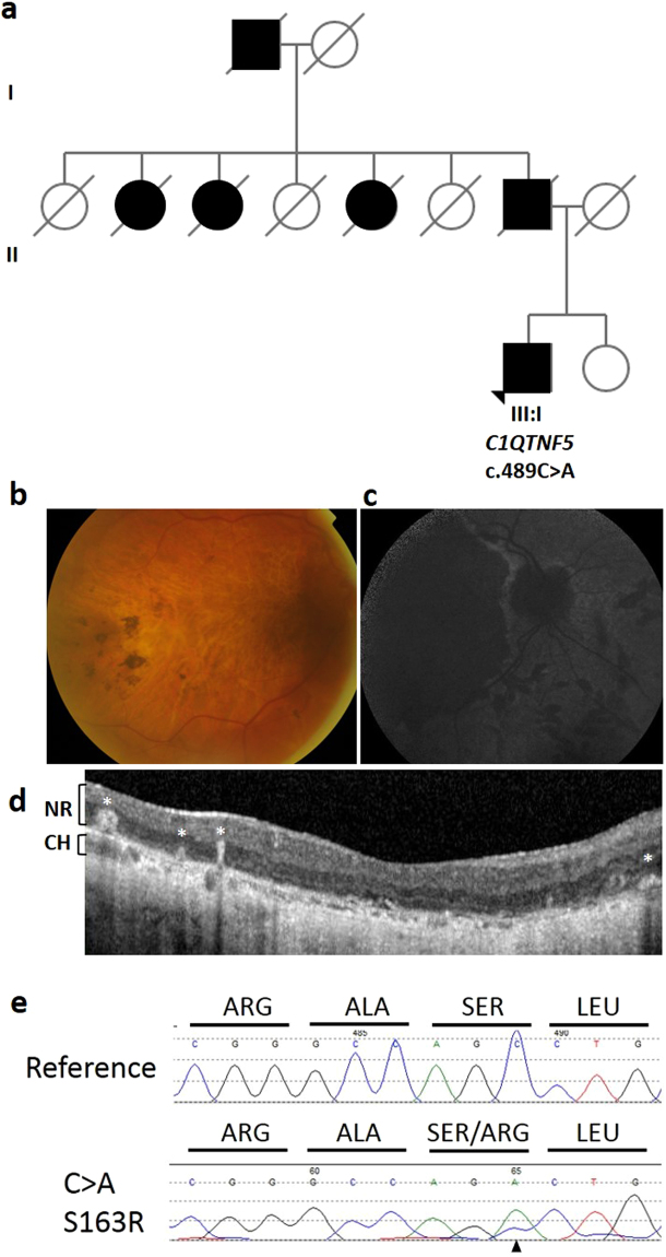
Pedigree, genotype and phenotype of family 3. (a) L-ORD family 3 is a multi-generation pedigree showing autosomal dominant inheritance. Squares represent males, circles represent females. Shaded shapes indicate an affected individual. Clinical phenotyping of the proband III:I (b) Colour fundus photography shows areas of central retinal atrophy. (c) Fundus autofluorescence imaging shows areas of hypoautofluorescence matching the areas of atrophy. (d) Hyperreflective deposits within and deep to the neural retina, indicated by white asterisks, are observed in the macula in an OCT scan of the proband. NR, neural retina; CH, choroid. (e) Sequence analysis of C1QTNF5 exon 3 revealed a heterozygous c.489 C > A transversion, resulting in a serine to arginine mutation at position 163 in the L-ORD case (lower panel) compared to an unrelated, unaffected individual (upper panel).

