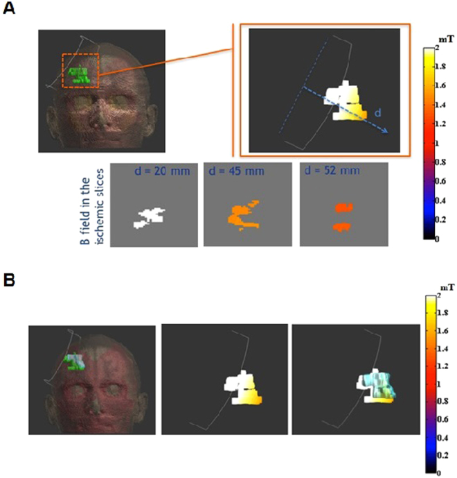
Figure 2.
Dosimetric results on exemplificative patient. (a) B field in ischemic lesion on the top and on slice perpendicular to the coil axis on the bottom; (b) on the left, geometrical comparison between the ischemic volumes before (green area) and after (light blue area) the magnetic field treatment, in the centre, B field on the pre-treatment injured volume; on the right, the same information overlapped with the geometric extension of the post-treatment area.

