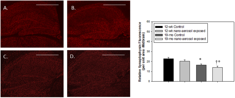
Figure 9.
Synaptophysin expression (red fluorescence) from the midbrain region of (A) 12–13 wk control; (B) 12–13 wk nano-aerosol-exposed; (C) 19 mo control; and (D) 19 mo nano-aerosol-exposed rats at 28 days after the end of exposure. A minimum of 3 locations on each section (2 sections per slide), 2 slides and n = 3 per group were analyzed for total fluorescence. Relative fluorescence per unit area is represented in the graph shown. *P < 0.050 compared to 12–13 wk control; †p < 0.050 compared to 19 mo control. Scale bar = 1000 µm.

