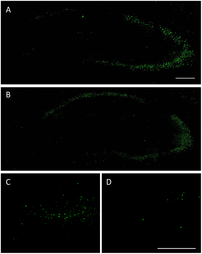
Figure 10.
Representative Fluoro-Jade C (FJC) stained coronal sections (section level −1.82 mm from bregma) of the hippocampal formation of the ipsilateral hippocampus of mice that received unilateral injection of kainate into the right CA1 at −2.1 mm from bregma one week before FJC staining. The section shown in “A” is from a vehicle control, while the section shown in “B” is from a drug treated mouse. “C” and “D” are enlarged views of the dentate hilus, illustrating the markedly reduced FJC staining in mice treated with NBQX and ifenprodil (D) compared to vehicle control (C). The scale bars in “A” and “D” indicate 200 µm.

