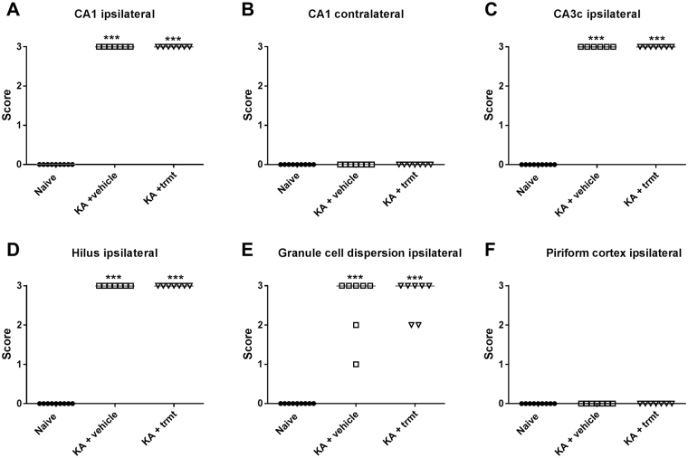
Figure 6.
Neurodegeneration following intrahippocampal kainate injection in mice. Data are from mice of groups 2 and 3, which were killed 8–22 weeks after kainate (see Fig. 1). A group of naive mice was used for comparison. Severe neurodegeneration was observed in the ipsilateral hippocampus in CA1 (A), CA3 (C) and dentate hilus (D), but not in the contralateral hippocampus (B) and ipsilateral (F) or contralateral piriform cortex. In addition to neuronal loss, granule cell dispersion was observed in the ipsilateral dentate gyrus (E). Individual data are shown; the group median is indicated by the horizontal line. Significant differences to naive controls are indicated by asterisk (P < 0.001). All data are from a section level of −1.94 mm from bregma (kainate was injected at −2.1).

