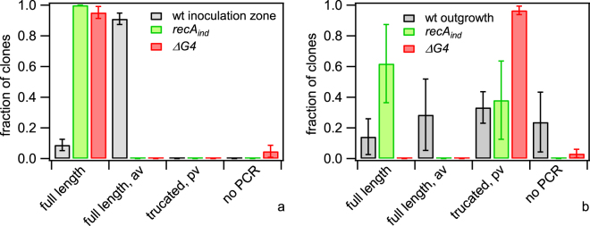
Figure 9.
Pilin variation in antigenic variation deficient strains. Cells were picked from (a) inoculation zone, and (b) outgrowth, respectively. After dilution and growth on agar plates, individual colonies were picked and pilE was sequenced. Sequences were categorised and the fraction shown is the number of sequences per category normalised by the total number of sequences found for each strain. Full length: most abundant sequence in inoculum; full length, av: sequence change mappable to pilS sequence; truncated, pv: length change in poly-C sequence causing premature stop-codon; no PCR: PCR amplification did not result in a product. Grey: red wt (Ng106) and green wt (Ng165), green: green recA (Ng167) and red recA (Ng168), red: green ∆G4 (Ng169) and red ΔG4 (Ng170). Mean and standard error of three independent experiments.

