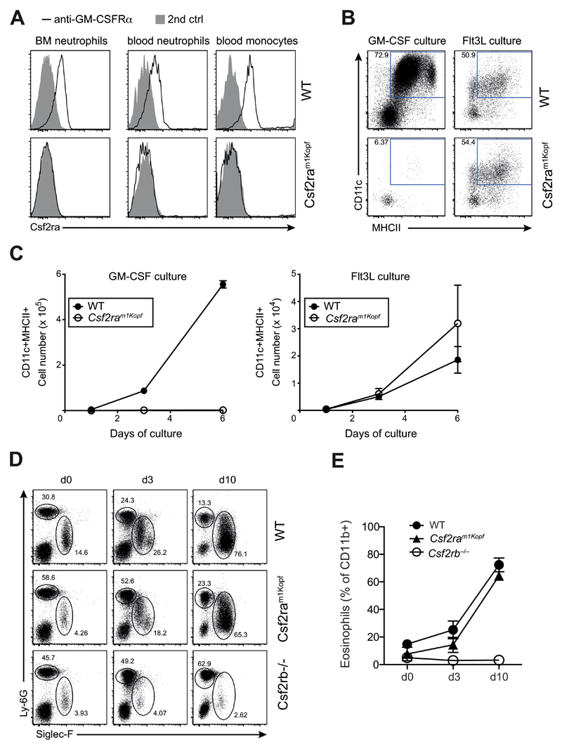
Figure 3. Csf2ram1Kopf mice are functional knockouts for the GM-CSFRα.
(A) Flow cytometry analysis of GM-CSFRα on neutrophils in the bone marrow (BM) or blood and monocytes in the blood of WT and Csf2ram1Kopf mice using anti-GM-CSFRα or secondary anti-ratIgG2a control. (B) In vitro differentiation of DCs from BM cells of mice as in (A) using GM-CSF or Flt3L. Expression of CD11c and MHCII on live cells is depicted. (C) Total numbers of CD11c+MHCII+ cells from in vitro DC cultures as in (B). (D) Flow cytometry of eosinophils and neutrophils in the blood after hydrodynamic injection of an IL-5 expression plasmid in WT, Csf2ram1Kopf and Csf2rb–/– mice. Numbers adjacent to outlined areas indicate percent Siglec-F+ eosinophils and Ly-6G+ neutrophils gated on CD11b+ cells. (E) Frequency of eosinophils as gated in (D).

