
Figure 2. Silencing of RORA attenuates hypoxia-induced expression of genes involved in late differentiation and epidermal barrier function in human keratinocytes.
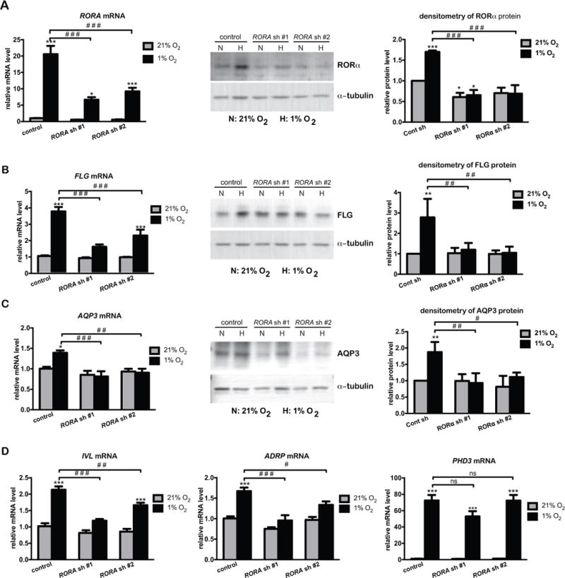
Real time RT-PCR analysis and western blot analysis of the expression of indicated genes. HaCat cells were stably transduced with lentivirus prepared from pLKO.1 vector (control) or two pLKO.1-RORA shRNAs. Cells were cultured under normoxic (21% O2) or hypoxic (1% O2) conditions for 24 h, and harvested for real time RT-PCR or western blot analysis of RORα (A), FLG (B), AQP3 (C), or other indicated genes (D). The mRNA level of each gene is normalized to 36B4. The protein level of indicated genes was quantified by densitometry scanning, and normalized to α-tubulin. Values are shown as mean-fold over control ± S.E.M. *, p < 0.05, **, p < 0.01 ***, p < 0.001, N=3 independent experiments.