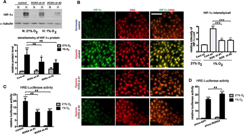
Figure 4. RORα is required for HIF-1α nuclear accumulation and transcriptional activity in human keratinocytes.

(A–C) HaCat cells stably transduced with lentivirus prepared from pLKO.1 vector (control) or two different pLKO.1-RORA shRNAs were cultured under normoxic (21% O2) or hypoxic (1% O2) conditions. (A) After 8-h culture under hypoxia, cells were harvested for western blot analysis of HIF-1α. (B) After 8-h culture under hypoxia, cells were fixed and subjected to immununostaining with an antibody against HIF-1α (green). DNA was counter stained with propidium iodide (PI). Bar = 70 μm. Fluorescence intensity of HIF-1α signal/cell was normalized to the intensity of DNA staining. Fifty cells from 10 independent fields were measured for each condition. Data are presented as mean-fold over control ± S.E.M. **, p < 0.01 ***, p < 0.001, N=3. (C) HaCat stable cells were transiently transfected with the pGL2-HRE-luciferase and renilla constructs. At 24 h post transfection, cells were cultured under normoxia or hypoxia for 24 h, and lysed for measurement of luciferase activities. (D) HaCat cells stably transduced with the retrovirus of pinco-GFP or pinco-RORα4 were transiently transfected with the pGL2-HRE-luciferase and renilla constructs, followed by the steps as described in (C). The HRE-luciferase activity was normalized to renilla activity, and presented as mean-fold over control ± S.E.M. **, p < 0.01 ***, p < 0.001, N=3.
