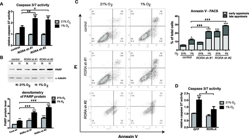
Figure 5. RORα protects keratinocytes during hypoxia.

(A–C) HaCat cells stably transduced with lentivirus prepared from pLKO.1 vector (control) or two different pLKO.1-RORA shRNAs were cultured under normoxic (21% O2) or hypoxic (1% O2) conditions. (A) After 24 h of normoxic or hypoxic culture, HaCat cells were analyzed for the caspase 3/7 activity, which was normalized to the number of viable cells measure by the Celltiter Glo assay. The value is presented as mean-fold over control ± S.E.M. N=3. (B) After 24 h of normoxic or hypoxic culture, cells were harvested for western blot analysis using an antibody against the cleaved form of PARP. The protein level of cleaved PARP was quantified as described in Fig. 1. (C) After 48 h of culture under normoxia or hypoxia, HaCat cells were double stained with Annexin V-FITC and propidium iodide (PI), and analyzed with flow cytometry for apoptotic cell death. Cells that stain positive for Annexin V-FITC and negative for PI are categorized as in the early apoptotic stage. Cells that stain positive for both Annexin V-FITC and PI are categorized as in the late apoptosis. Values show mean-percentage of total cells ± S.E.M., N=3 independent experiments. (D) HaCat cells stably transduced with the retrovirus expressing GFP or RORα4 were culture under normoxia or hypoxia for 24 h, and analyzed for the caspase 3/7 activity, as described in (A).
