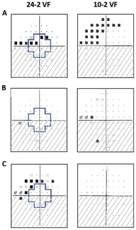
Figure.
The region within the blue outline on the 24-2 visual fields corresponds to the region in the central 10° tested by the 10-2 visual field test points. All fields are presented in right eye view. (A) Both superior hemifields are abnormal. (B) The superior hemifield is normal on the 24-2, but abnormal on the 10-2. (C) The superior hemifield is abnormal on the 24-2, but normal on the 10-2. Source: Traynis I, De Moraes CG, Raza AS, et al. JAMA Ophthalmology. 2014;132(3):291–297.4

