
Figure 6. Patch clamp recordings of apical K channels in the TAL of mice on CTRL and LNaHK.
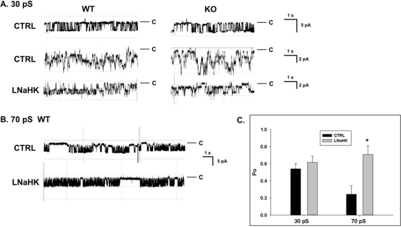
A. Recordings of single (top; CTRL, −Vp = −60 mV) and multiple (−Vp = −40 mV) 30 pS (inward currents) K channels from cell-attached patches of split-open TAL of WT and KO mice on CTRL or LNaHK. Pipette solution contained 140 mM KCl. Red lines denote closed states. B. Recordings of 70 pS Kir channels in apical membrane of WT mice. The recordings indicate 2 closed states (long and short) with an increased duration in long closed state in channels of mice on CTRL. C. Summary of Po at −Vp = −40 mV of the 30 pS and 70 pS Kir in the TAL from WT on CTRL and LNaHK. *p < 0.05 vs CTRL analyzed by one-way ANOVA.