
Figure 7. ROMK localization in the TAL of mice on CTRL and LNaHK.
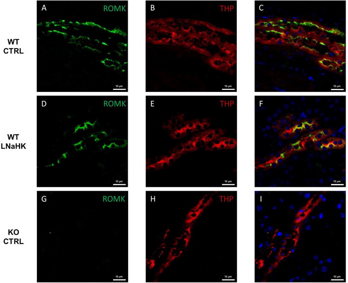
A-C. Immunostaining of ROMK (green), Tamm-Horsfall Protein (THP; marker of TAL; red), and both in the outer medulla of WT on CTRL. D-F. Immunostaining of ROMK (green), THP (red), and both in the outer medulla of WT on LNaHK. G-I. Immunostaining of ROMK (green), THP (red), and both in the outer medulla of KO on CTRL. All images were taken at 400× magnification. All scale bars are 10 μm.