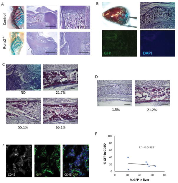
Figure 4. Rescue of bone marrow hematopoiesis by stem cell-derived osteoblasts.
(A) Skeletal preparations and hematoxylin- and eosin-stained sections of the proximal humerus of E18.5 control (upper) and Runx2−/− (lower) embryos. Note the absence of hematopoietic bone marrow in the Runx2−/− humerus. Scale bar = 1 mm (middle panels) or 200 μm (right panels). (B) Skull skeletal preparation demonstrating partial rescue in a Runx2−/− host blastocyst injected with stem cells. Histological analysis reveals the formation of a bone marrow cavity. GFP expression confirms chimeric contribution of stem cells. Scale bar = 200 μm. (C) Histological analyses of humeri of Runx2−/− blastocysts injected with ES cells. ES contribution (%) as determined by flow cytometry is provided under each panel. Scale bar = 200 μm. (D) Histological analyses of humeri of Runx2−/− blastocysts injected with iPS cells. iPS contribution (%) as determined by flow cytometry is provided under each panel. Scale bar = 200 μm. (E) Immunohistochemical staining for CD45 (white), GFP (green) and nuclei (DAPI, blue) reveals CD45+ hematopoietic cells in the bone marrow. (F) The frequency of CD45+ hematopoietic cells that are stem cell-derived (GFP+) does not correlate with the % GFP contribution in the liver.

