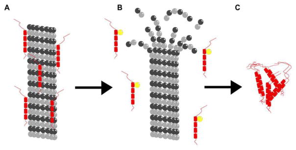
Figure 1.
Schematic illustration of microtubule depolymerization in Alzheimer’s disease neurons. (A) Microtubule (gray) stabilized by tau (red). (B) Destabilized microtubule and free tau with additional phosphorous (yellow). (C) Neurofibrillary tangles formed from phosphorylated tau. This image is a reproduction of the original found in Ref. [70] and presented under the PLoS One Creative Commons Attribution 4.0 International Public License (CC BY 4.0).

