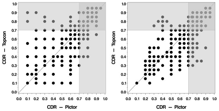
Figure 1.
Initial cup to disc ratio (CDR) measurements obtained from the Topcon versus the Pictor images of the posterior pole, stratified by grader (Left. Grader 1; Right. Grader 2). The light gray shaded area indicates where the CDR is measured above the threshold for a diagnosis of glaucoma (≥0.7) by one camera but not the other.

