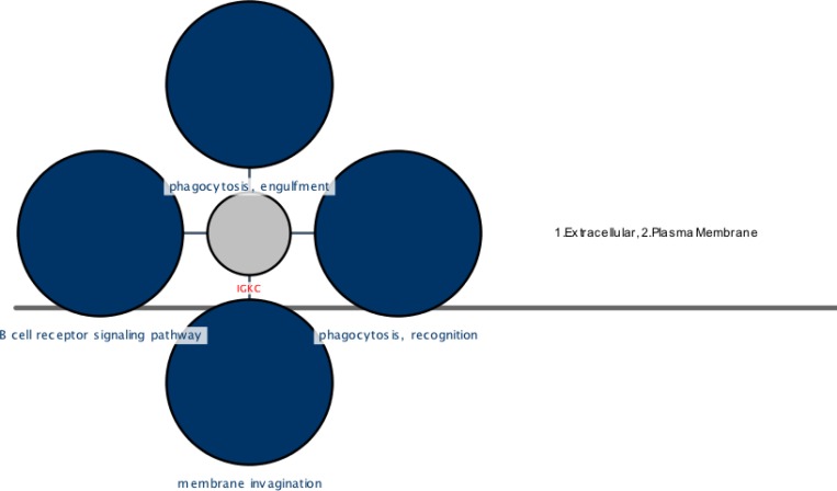
Figure.3.
A cerebral view of the IGKC showing cellular locations and their related four top ranked biological processes (BP) colored in blue. These parts are coded as 1 (extracellular), 2 (plasma membrane) for IGKC. The phagocytosis, recognition, phagocytosis, engulfment, membrane invagination, and B cell receptor signaling pathway are the processes shown as dark blue circles and extracellular and plasma membrane are the locations of IGKC that are mentioned in the right position of the figure. The pvalue< 0.05

