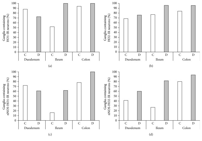
Figure 4.
Percentage of myenteric ganglia containing HO1-IR (a), HO2-IR (b), nNOS-HO1-IR (c), and nNOS-HO2-IR (d) neurons. Only half of the ileal ganglia contained HO1-IR neurons, and from these only 16% of the ganglia represented double-stained neurons in the control rats. In the diabetic group, all of the ileal and colonic ganglia included HO1-IR neurons, and from these more than 60% of ileal and 100% of colonic ganglia also represented the double-stained neurons. More than 70% of ganglia represented HO2-IR neurons under both experimental conditions, and a large increase in the proportion of ganglia containing double-stained neurons was revealed in diabetic state. C: controls; D: diabetics.

