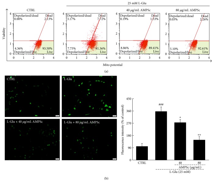
Figure 3.
(a) The dissipation of MMP caused by 12 h L-Glu incubation was strongly restored by 3 h AMPSc pretreatment analyzing via JC-1 staining (n = 6). (b) The overproduction of ROS induced by 12 h L-Glu exposure was significantly decreased by 3 h AMPSc pretreatment analyzed by DCFH-DA staining (n = 6). Scale bar: 20 μm. Qualification data were expressed as the percentage of green fluorescent intensity compared to control cells. Data are expressed as means ± S.E.M. (n = 6). ###P < 0.001 versus CTRL; ∗P < 0.05 and ∗∗P < 0.01 versus L-Glu-exposed cells. AMPS: A. mellea polysaccharides.

