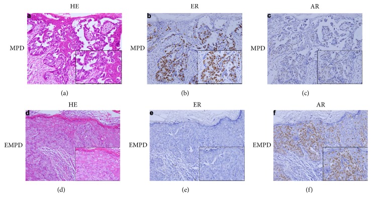
Figure 1.
Representative images of histological and immunohistological features of MPD and EMPD. (a) HE staining of MPD, (b) nuclear positive staining of ER in MPD, (c) nuclear negative staining of AR in MPD, (d) HE staining of EMPD, (e) negative staining of ER in EMPD, and (f) nuclear positive staining of AR in EMPD.

