
Fig. 2.
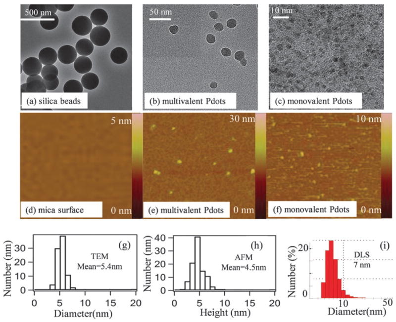
Size distribution of mPdots measured using TEM, AFM, and DLS. (a) TEMimage of silica beads showing a diameter of ~200 nm. (b) TEM image of regular PPV–PPA Pdots, which have an average diameter of 34 ± 4 nm, from measurements on 80 Pdots. (c) TEM image of PPV–PPA mPdots. (d) AFM image (1 μm × 1 μm) of the 3-aminopropyltriethoxysilane (APTEOS)-coated mica surfacewithout any nanoparticles. (e) AFM image (0.6 μm × 0.6 μm) of multivalent regular PPV–PPA Pdots. (f) AFM image (1.2 μm × 1.2 μm) of PPV–PPA mPdots. (g) Size distribution of mPdots shown in (c); the average diameter was 5.4 ± 0.5 nmfromimages of 88 mPdots. (h) Height distribution of mPdots measured using AFM; the average value was 4.5 ± 0.4 nm from measurements on 100 mPdots. (i) DLS results of mPdots in aqueous solution showing a hydrodynamic diameter of 7 nm.