
Figure 4.
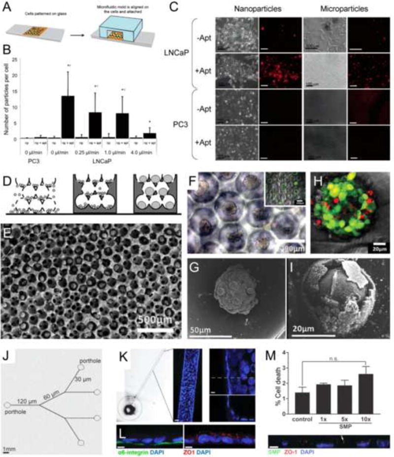
Cancer-on-a-chip platforms for evaluating nanomedicine. (a–c) A microfluidic planar culture model for evaluating nanoparticle (NP) interactions with cancer cells. (a) Schematic of the device fabrication. (b,c) Quantification and images showing the uptake of prostate-specific membrane antigen (PSMA)-functionalized poly(lactic acid) (PLA) nano- and microparticles by PC3 and LNCaP prostate cancer cells cultured in the microfluidic device. (d–i) An inverted opal 3D cancer model for evaluating NP interactions with cancer cells. (d) Schematic of the model fabrication. (E–G) SEM and optical microscopy images showing the liver cancer spheroids formed in the inverse opal scaffolds. Inset in (f) shows the viability of the spheroids. (h,i) Viability analysis and SEM image showing the liver cancer spheroids after treatment with CdTe semiconductor NPs. (j–l) A biomimetic mammary duct-on-a-chip platform. (j) Schematic of the branching microchannel system built in a polydimethylsiloxane (PDMS) chip. (k) Formation of mammary epithelium surrounding the periphery of the microchannel. (l) Confocal micrographs showing staining for both basal (α6 integrin) and apical (ZO-1) polarity markers. (m) Dose-dependent cell death caused by injected submicron magnetic particles (SMPs); the bottom panel shows a confocal image of the co-registration of the SMPs with the epithelium. Adapted, with permission, from [56] (b,c), [57] (h,i),and [58] (m).