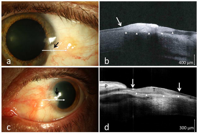
Figure 2.
(a) A 37-year-old male boat captain with a longstanding conjunctival lesion presented with a subtle area of leukoplakia (black arrow) at the corneal margin of the lesion. (b) HR-OCT image (commercial device, white line scan in a) demonstrated hyper-reflective thickened epithelium (above arrow heads) and an abrupt transition zone (white arrow). (c) Similarly, a subtle opacification and elevation was noted at the head of a longstanding pterygium of a 64-year-old male (black arrow). (d) HR-OCT (commercial device, white line scan in c) revealed the anatomy of the folded pterygium tissue (P) with normal epithelium (E), and an adjacent area of thickened epithelium (above arrow heads, T), with two abrupt transition zones (white arrows) suspicious for OSSN. Biopsy confirmed OSSN in both cases.

