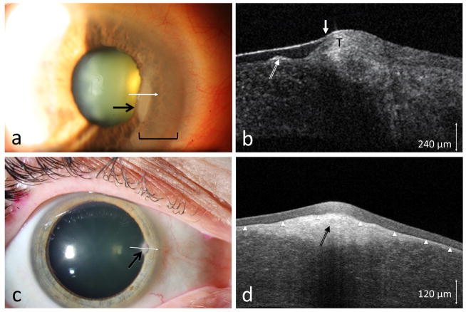
Figure 3.
(a) A 76-year-old male seen with an opacity (area within bracket) adjacent to a long standing nodule (black arrow). (b) HR-OCT (custom device, white line scan in a) showed the subepithelial opacities (long white arrow) consistent with Salzmann’s nodular changes but also subtle adjacent epithelial thickening (T) and a transition zone (short white arrow) consistent with OSSN. Biopsy confirmed OSSN. (c) A 57-year-old female referred for a corneal opacity thought to be OSSN (black arrow). (d) HR-OCT (commercial device, white line scan in c) demonstrated thin, dark epithelium (above the arrowheads) with a hyper-reflective subepithelial lesion (black arrow) consistent with Salzmann’s, which was confirmed by biopsy.

