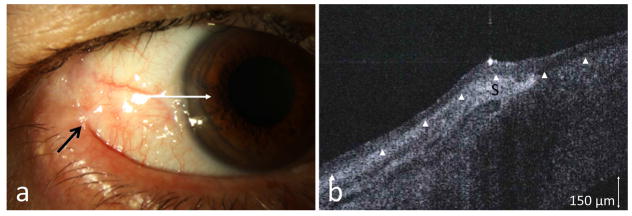
Figure 4.
(a) A 73-year-old male with a history of OSSN in the lateral canthus 7 years prior to presentation developed Stage III MMP (Foster classification) with symblepharon formation (black arrow) and presented for possible OSSN recurrence at the limbus. (b) HR-OCT (commercial device) along the white line, however, demonstrated thin epithelium (above arrowheads) with a subepithelial hyper reflectivity consistent with scarring (S) and no OSSN. Biopsy was negative for OSSN.

