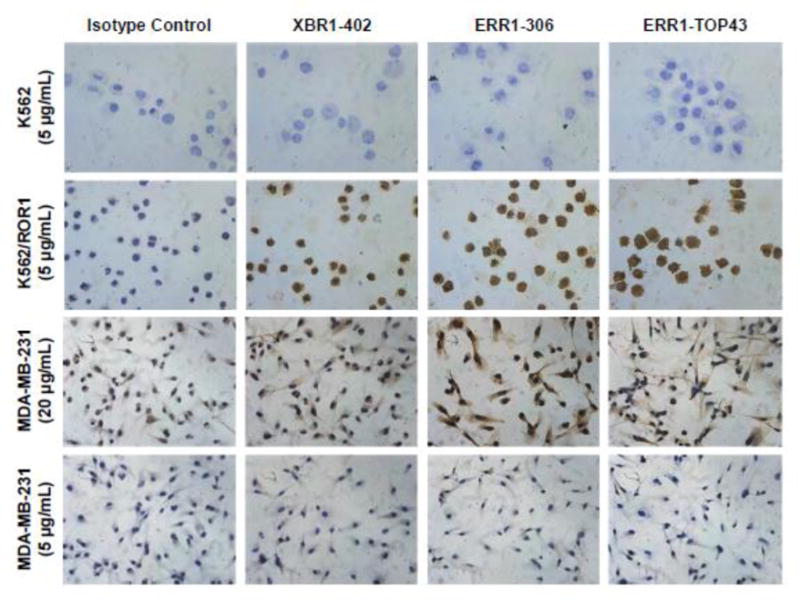
Figure 4. Analysis of selected full rabbit anti-human ROR1 IgG1s by IHC.

Ectopically expressed ROR1 (K562/ROR1 vs. K562 cells) and endogenously expressed ROR1 (MDA-MB-231 cells) was detected by IHC using full rabbit IgG that were generated from XBR1-402, ERR1-316, and ERR1-TOP43. Note that the IHC detection of endogenously expressed ROR1 on the cell surface of fixed human cells and tissues by mAbs to the ECD of ROR1 is known to be difficult.58 Fittingly, a high concentration of rabbit mAbs (20 μg/mL) was required for staining the MDA-MB-231 cells. All light microscopy images were taken at a magnification of 40× (total magnification of 400×).
