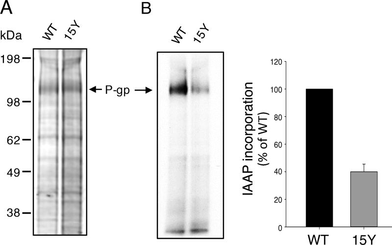
Fig. 6. 15Y mutant P-gp exhibits decreased incorporation of photoaffinity substrate IAAP.
Equal amounts of high five insect cell membranes expressing either WT or 15Y mutant P-gp (75 µg protein) were photo-crosslinked with IAAP (5–7 nM) as described in Materials and Methods. After 5 minutes, the samples were exposed to UV light for 10 min to crosslink bound IAAP to Pgp. Then the samples were prepared for electrophoresis. Labeled 30 µg protein was loaded per lane and the gels were stained for protein with colloidal blue dye, dried and scanned. Representative protein stained gel in (A) and an autoradiogram of the same gel in (B) is shown. The incorporation of IAAP in WT and 15 Y mutant P-gp was normalized to P-gp level in the stained gel (A) from three independent experiments (mean ± SD; bar graph on the right).

