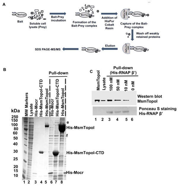
Figure 2. Pull-down assay of MsmTopoI-RNAP interaction with HisPur Cobalt agarose resin.
(A) General scheme of the pull-down assay. (B) Proteins pulled down from M. smemgatis soluble lysate by recombinant N-terminal His-tagged MsmTopoI (lane 8) or MsmTopoI-CTD (lane 7). His-Mocr, a recombinant viral protein, was used as bait in the control reaction (lane 6). Lane 1: MW standards; Lane 2: purified recombinant His-Mocr; Lane 3: purified recombinant His-MsmTopoI-CTD; Lane 4: purified recombinant His-MsmTopoI. (C) Reverse pull-down of TopoI from M. smegmatis soluble lysate with purified recombinant RNAP β′ subunit (N-terminal His-tagged). Following SDS-PAGE, the proteins eluted from the HisPur Cobalt were analyzed by western blot using anti-TopoI antibodies. Lane 1: purified recombinant MsmTopoI (100 ng); Lane 2: 15 μg of total proteins in M. smegmatis soluble lysate; Lanes 3–6: eluates from pull-down assay using 500 μg of total proteins in M. smegmatis soluble lysate with His-tagged RNAP β′ subunit of concentrations 100 nM (lane 3), 50 nM (lane 4), 10 nM (lane 5), and 0 nM (lane 6). The nitrocellulose membrane was also stained with Ponceau S for detection of the recombinant bait (His-tagged RNAP β′) used in the assay.

