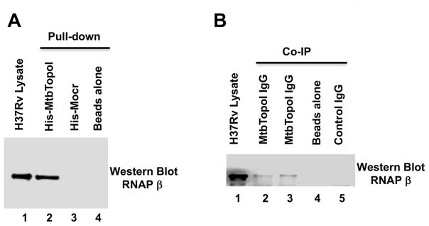
Figure 3. M. tuberculosis H37Rv topoisomerase I and RNAP are protein-protein interaction partners.
M. tuberculosis RNAP was detected by western blot using a monoclonal antibody against E. coli RNAP β subunit that cross-reacts with mycobacterial RNAP. (A) Pull-down assay. Lane 1: total soluble lysate of M. tuberculosis H37Rv (10 μg). Eluates from HisPur cobalt resin incubated with 250 μg of lysate and purified His-tagged MtbTopoI (lane 2), 6xHisMocr (lane 3) or without any His-tagged protein (lane 4). (B) Co-Immunoprecipitation assay. Lane 1: total soluble lysate of M. tuberculosis H37Rv (10 μg). Immunoprecipitates from 250 μg of total soluble lysates incubated with IgG against MtbTopoI (lanes 2, 3), no IgG added (lane 4), and IgG from pre-immune serum (lane 5) were analyzed by western blot with the antibody recognizing RNAP β subunit.

