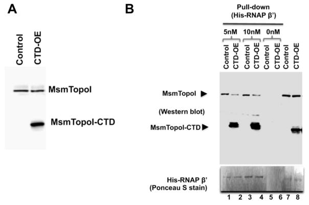
Figure 6. Inhibition of MsmTopoI-RNAP interaction with overexpression of recombinant MsmTopoI-CTD.
(A) The tetracycline-induced overexpression of MsmTopoI-CTD was confirmed by western blot analysis with rabbit polyclonal antibodies against TopoI. Lane 1: lysate (10 μg) of M. smegmatis transformed with control vector. Lane 2: lysate (10 μg) of M. smegmatis transformed with pMsmTopoI-CTD. (B) Pull-down of MsmTopoI from M. smegmatis lysate by His-tagged RNAP β′ subunit is reduced by the competing overexpressed MsmTopoI-CTD. Pull-down of MsmTopoI and MsmtopoI-CTD from the lysate (350 μg) by His-RNAP β′ (5 nM or 10 nM) was analyzed by western blot using antibodies against MtbTopoI (upper panel). Lanes 1, 3: pull-down reactions with lysate from the strain transformed with the control vector (control) in the presence of 5 nM His-RNAP β′ or 10 nM His-RNAP β′. Lanes 2, 4: pull-down reactions with lysate from the strain overexpressing the MsmTopoI-CTD (CTD-OE) in the presence of 5 nM His-RNAP β′ or 10 nM His-RNAP β′. Lanes 5, 6: lysates from either the control strain or the overexpression strain were incubated with the beads as a negative control for the pull-down assays. Lane 7: lysate (10 μg) from M. smegmatis transformed with the control vector. Lane 8: lysate (10 μg) from M. smegmatis overexpressing the MsmTopoI-CTD. The nitrocellulose membrane was stained with Poneau S (bottom panel) for the detection of the bait (His-RNAP β′).

