
Figure 2.
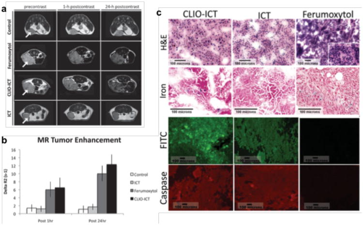
(a) Axial T2-weighted MR images (TR 2500 ms, TE 80 ms) of MMTV-PyMT mammary tumors before and after a single intravenous injection of different components. (b) MR signal enhancement data in tumors quantified as ΔR2 = (R2pre - R2post). (c) Cell death in MMTV-PyMT tumors. H&E panels: CLIO-ICT treated tumor demonstrating diffuse necrosis; ICT treated tumor with predominately viable tumor cells and a subset of cells undergoing necrosis; Ferumoxytol treated tumor with diffuse viability and no necrosis. Iron panels: Scattered CLIO-ICT treated tumor and rare admixed histiocytes contain blue pigment indicating cytoplasmic iron deposition; ICT treated tumor shows no cytoplasmic iron deposition, scattered iron laden histiocytes serve as an internal positive control; Ferumoxytol treated tumor show cytoplasmic iron deposition, scattered iron laden histiocytiocytes serve as an internal positive control. FITC panels: Fluorescence microscopy showing FITC signal for CLIO-ICT and ICT but no signal for Ferumoxytol. Caspase-3 panels: CLIO-ICT and ICT treated tumors show Cy3 labeling throughout the samples; Ferumoxytol treated tumor shows few areas with weak Cy3 fluorescence. Reprinted with permission from WILEY-VCH Verlag GmbH & Co. KGaA, Weinheim [32].