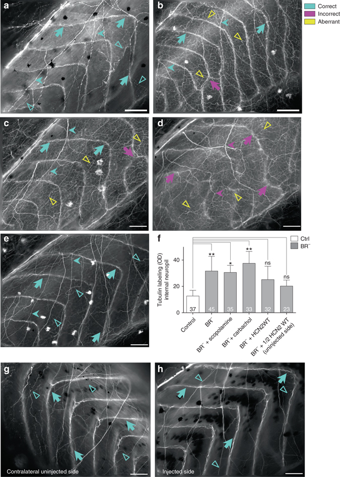
Fig. 5.

The absence of a brain generates an abnormal neural network in the entire animal body. a, b Acetylated-tubulin (Tub) immunoexpression for Ctrl (a) and BR− (b) animals. There three types of nerve fibers: (i) commissural fibers (dorsoventral axis, long arrows); (ii) longitudinal fibers (anteroposterior axis, short arrow); and (iii) internal neuropil (no defined axis, unfilled triangles). Animals developed without a brain show normal commissural and longitudinal nerve fibers (turquoise long arrows in b), with some alterations (magenta long arrow), but a dense internal neuropil (yellow unfilled triangles). c, d Tub-immunoexpression for BR− treated with cholinergic drugs: scopolamine (c) and carbachol (d). Scopolamine treatment was not able to rescue the aberrant internal network (yellow unfilled triangles in c), and carbachol-treated animals exhibited a chaotic nerve patterning (magenta and yellow arrows in d). e Ectopic HCN2-WT expression (injected in both cells at two-cell stage) fixed the BR−–induced internal nerve branching. f Quantification of the mean OD of internal neuropil and statistical comparisons among untreated/uninjected Ctrl and untreated/uninjecetd BR− (BR−, without drug treatment nor ion channel misexpression), scopolamine-treated BR− (BR− + scopolamine), carbachol-treated BR− (BR− + carbachol), HCN2-WT both-sides injected BR− (BR− + HCN2 WT), and HCN2-WT LR side-injected BR− (BR− + 1/2 HCN2 WT) embryos (one-way ANOVA, P < 0.01). No significant differences after a posteriori analysis were detected among the different Ctrl groups. Data represent the mean OD units and s.d. of two independent replicates. Number in bars indicates n or number of animals analyzed for each group. P values after post-hoc Bonferroni’s test are indicated as **P < 0.01, *P < 0.05, ns P > 0.05. g, h. Ectopic HCN2 expression in only one LR side fixes the BR−-induced internal nerve branching. Tub-immunoexpression on β-gal-reacted sections (dark deposits) in a 1/2 HCN2-WT BR−, showing both the contralateral uninjected side (g) and the injected (h) of the same embryo. Aberrant neural network was completely rescued (turquoise arrows), exhibiting a similar nerve pattern to the Ctrl group in both sides. a-e, h: Rostral is upper right and dorsal is up. g: Rostral is upper left and dorsal is up. Scale bar, 100 μm
