
Figure 7. Effects of deficiency of Ccr2, Cx3cr1 or both on metabolic and inflammatory phenotypes in female mice.
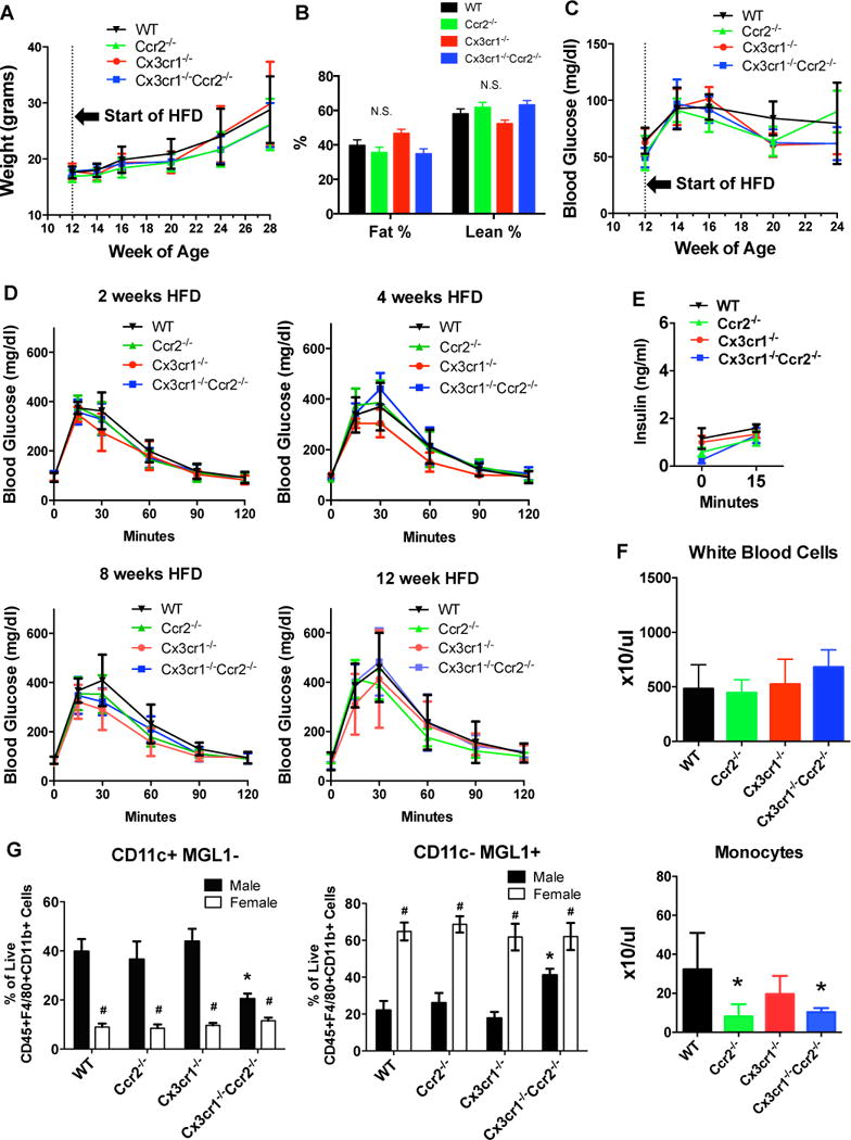
Female WT, Ccr2−/−, Cx3cr1−/− and Cx3cr1−/−Ccr2−/− mice were fed a 45% HFD starting at 12-weeks of age (n=10 WT, 10 Ccr2−/−, 5 Cx3cr1−/−, 5 Cx3cr1−/−Ccr2−/−). A: Body weight was measured every 2–4 week. B: Body composition was measured by NMR after 21 weeks of HFD. C and D: Fasting blood glucose and IPGTT were examined in female mice using the same protocols as in male mice. E: Glucose-induced insulin secretion was measured after 13 weeks of HFD after an overnight fast and was not different between groups. F: Blood count with differential was performed after 19 weeks of HFD. *, P<0.05 compared with female WT mice. G: Flow cytometric analysis of stromal vascular fraction of perigonadal adipose tissue was performed in female mice and data were compared with male mice. *, P<0.05 when comparing male WT mice with male mice of other genotypes; #, P<0.05 when comparing male and females mice of the same genotype. Data are mean±SEM. n=6 female WT, 6 female Ccr2−/−, 4 female Cx3cr1−/−, 5 female Cx3cr1−/−Ccr2−/−. N.S., not significant.