Figure 3.
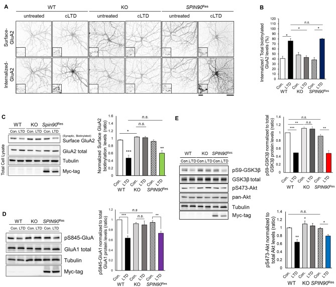
Chemical forms of LTD (cLTD)-induced AMPAR endocytosis is blocked in Spin90-KO neurons. (A) AMPAR internalization assay on DIV18 hippocampal neurons. Surface GluA2 levels indicate constitutive AMPAR trafficking and internalized levels are those endocytosed after cLTD induction (100 μM NMDA application for 5 min and 15 min wash). Scale bar, 5 μm and 20 μm. (B) Internalized GluA2 levels were plotted against total biotinylated GluA2 and quantified (n = 8–15 for WT and Spin90-KO; n = 3 for adeno-associated virus (AAV)-6xMyc-SPIN90 infected Spin90-KO neurons (SPIN90Res). (C) Immunoblot analysis of surface biotinylated GluA2 levels after cLTD induction in WT, Spin90-KO and SPIN90Res neurons (n = 4 for all genotypes). (D) Immunoblot analysis of dephosphorylation of Ser845-GluA1 after cLTD induction (n = 9 for WT and Spin90-KO; n = 4 for SPIN90Res). (E) Changes in phosphorylation of Akt-GSK3β signaling upon cLTD induction (n = 14 for WT and Spin90-KO; n = 4 for SPIN90Res). All data are expressed as mean ± SEM (*p < 0.05, **p < 0.01, ***p < 0.001, n.s., non-significant).

