Figure 5.
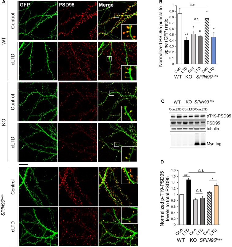
SPIN90 is necessary for PSD95 dissociation during cLTD. (A) DIV18–21 WT, Spin90-KO and AAV-6xMyc-SPIN90 infected Spin90-KO neurons (SPIN90Res) hippocampal neurons expressing GFP and PSD95 were pre-incubated with CNQX (10 μM) for 1 h before cLTD treatment, followed by immunostaining with GFP (green) and PSD95 (red) antibody. Scale bars indicate 5 μm. (B) The localization of PSD95 in ratios of PSD95 to GFP puncta was analyzed (n = 13–21 for WT and Spin90-KO; n = 19 for SPIN90Res-control(Con), n = 21 for SPIN90Res-LTD). (C) DIV18 WT, Spin90-KO and SPIN90Res neurons were subjected to cLTD treatment followed by harvesting of cells and immunoblot analysis. Phosphorylation of PSD95 at Thr19 was assessed. (D) Quantification of pThr19-PSD95 levels normalized to total PSD95 after cLTD induction (n = 9 for WT and Spin90-KO; n = 3 for SPIN90Res). All data are expressed as mean ± SEM (#p = 0.0629, *p < 0.05, **p < 0.01, n.s., non-significant).

