
FIGURE 7.
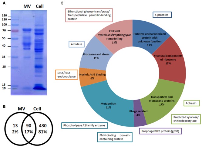
Microvesicles (MVs) are enriched in specific proteins. (A) Differential patterns observed between MV and cell extract proteins from L. casei BL23 in a Coomassie blue stained SDS-PAGE gel (n = 5). (B) Venn diagram of the number of proteins present in the MVs and cell extracts identified by LC-MS/MS. (C) Proteins of MVs identified by LC-MS/MS were grouped in 8 categories according to its function. The function of the 13 proteins exclusively present in the MVs are shown in boxes.